| Citation: |
WANG Qian, SUN Quanya, HE Min, ZHANG Shuo, XIANG Boni, LI Qiufan, WANG Yong, ZHANG Xialing, DING Tianling, YE Hongying. Clinical Characteristics and Diagnostic Experience of Adult Thyroid Langerhans Cell Histiocytosis with Diabetes Insipidus[J]. Journal of Rare Diseases, 2023, 2(3): 346-352. DOI: 10.12376/j.issn.2097-0501.2023.03.005

|
To analyze the clinical characteristics of thyroid LCH to enhance understanding of the disease.
We retrospectively studied the clinical data from six thyroid LCH patients who hospitalized in Huashan Hospital Affiliated to Fudan University from January 2015 to January 2022.We analyzed the ultrasound and 18F FDG-PET/CT imaging characteristics of thyroid LCH.
The six patients diagnosed (2 males and 4 females) were between 18 and 58 years old.All patients had diabetes insipidus.MRI revealed thickened pituitary stalk.Two cases had central hypothyroidism, while four cases euthyroidism.Three cases tested positive for thyroid antibodies.Ultrasound showed thyroid nodules of TI-RADS 3 in three cases, TI-RADS 4 in two cases, and 1 with nodular goiter.Ultrasound showed that all sic cases indicated low echogenicity, 5 of which clear boundaries, 4 of which uneven echo distribution, 5 of which irregular shape, and noen has calcification.18F FDG-PET/CT indicated high uptake nodules with SUVmax values all above 10.4 cases were diagnosed by surgical excision and the other 2 by coarse-needle aspiration biopsy.When diagnosed, two cases had liver and thymus involvement respectively.One case had lung and bone involvement respectively.After treatment, 4 cases showed that nodular goiter shrank, while the other two with liver involvement progressed fast and no assessment made.
Thyroid LCH presented low echogenicity, clear boundaries, irregular shape, without calcification, and high uptake in 18F FDG-PET/CT.A definite diagnosis of pituitary stalk thicking accompanied by thyroid nodules, especially those with hypoechoic and irregular nodules, can be achieved by coarse-needle aspiration biopsy and langerin-specific pathological staining.
| [1] |
Allen CE, Merad M, McClain KL. Langerhans-cell histiocytosis[J]. N Engl J Med, 2018, 379(9): 856-868. doi: 10.1056/NEJMra1607548
|
| [2] |
Goyal G, Tazi A, Go RS, et al. International expert consensus recommendations for the diagnosis and treatment of Langerhans cell histiocytosis in adults[J]. Blood, 2022, 139(17): 2601-2621. doi: 10.1182/blood.2021014343
|
| [3] |
Kwak JY, Han KH, Yoon JH, et al. Thyroid imaging reporting and data system for US features of nodules: a step in establishing better stratification of cancer risk[J]. Radiology, 2011, 260(3): 892-899. doi: 10.1148/radiol.11110206
|
| [4] |
Girschikofsky M, Arico M, Castillo D, et al. Management of adult patients with Langerhans cell histiocytosis: recommendations from an expert panel on behalf of Euro-Histio-Net[J]. Orphanet J Rare Dis, 2013, 8: 72. doi: 10.1186/1750-1172-8-72
|
| [5] |
吴蔚, 姚振威, 王镛斐, 等. 垂体柄增粗相关疾病——上海华山医院诊疗经验[J]. 中华内分泌代谢杂志, 2020, 36(7): 569-571. doi: 10.3760/cma.j.cn311282-20200306-00134
|
| [6] |
Cai HC, Liu T, Cai H, et al. Adult Langerhans cell histiocytosis with thyroid gland involvement: clinical presentation, genomic analysis, and outcome[J]. Ann Hematol, 2022, 101(9): 1925-1929. doi: 10.1007/s00277-022-04894-9
|
| [7] |
Patten DK, Wani Z, Tolley N. Solitary Langerhans histiocytosis of the thyroid gland: a case report and literature review[J]. Head Neck Pathol, 2012, 6(2): 279-289. doi: 10.1007/s12105-011-0321-8
|
| [8] |
陈海萍, 雷志锴, 李巧云, 等. 多系统朗格汉斯细胞组织细胞增生症一例并文献复习[J]. 中华医学超声杂志(电子版), 2019, 16(10): 794-797. doi: 10.3877/cma.j.issn.1672-6448.2019.10.015
|
| [9] |
Chen ED, Cheng P, Cai YF, et al. Ultrasonographic features of Langerhans cell histiocytosis of the thyroid[J]. Int J Clin Exp Pathol, 2014, 7(3): 1229-1235.
|
| [10] |
de Koster EJ, Noortman WA, Mostert JM, et al. Quantitative classification and radiomics of[18F]FDG-PET/CT in indeterminate thyroid nodules[J]. Eur J Nucl Med Mol Imaging, 2022, 49(7): 2174-2188. doi: 10.1007/s00259-022-05712-0
|
| [11] |
Cai YF, Wang QX, Ni CJ, et al. A case report: the diagnosis and therapeutic evaluation for a rare disease of Langerhans cell histiocytosis involving thyroid[J]. Medicine(Baltimore), 2015, 94(44): e1891.
|
| [12] |
Long Q, Shaoyan W, Hui W. 18F-fluorodeoxyglucose posi-tron emission tomography/computed tomography for primary thyroid Langerhans histiocytosis: a case report and literature review[J]. Indian J Nucl Med, 2015, 30(4): 328-330. doi: 10.4103/0972-3919.159688
|
| [13] |
Luo ZH, Lu PX, Qi WL, et al. Role of 18F-FDG PET/CT in the diagnosis and management of patients with Langerhans cell histiocytosis[J]. Quant Imaging Med Surg, 2022, 12(6): 3351-3363. doi: 10.21037/qims-21-823
|
| [14] |
Giovanella L, Ceriani L, Crippa S, et al. Imaging in endocrinology: Langherans cell histiocytosis of the thyroid gland detected by 18FDG-PET/CT[J]. J Clin Endocrinol Metab, 2007, 92(8): 2866-2867. doi: 10.1210/jc.2007-0336
|
| [15] |
Chen DW, Lang BHH, McLeod DSA, et al. Thyroid cancer[J]. Lancet, 2023, 401(10387): 1531-1544. doi: 10.1016/S0140-6736(23)00020-X
|
| [16] |
Zhu H, Hu DX. Langerhans cell histiocytosis of the thyroid diagnosed by fine needle aspiration cytology. A case report[J]. Acta Cytol, 2004, 48(2): 278-280. doi: 10.1159/000326332
|
| [17] |
Maraqa B, Al-Ashhab M, Kamal N, et al. Concomitant Langerhans cell histiocytosis of cervical lymph nodes in adult patients with papillary thyroid carcinoma: a report of two cases and review of the literature[J]. Autops Case Rep, 2021, 11: e2021253. doi: 10.4322/acr.2021.253
|

Official account

Supported by: Beijing Renhe Information Technology Co., Ltd. 
 Author
 Peer Review
 Editorial Entry
 Editor Work
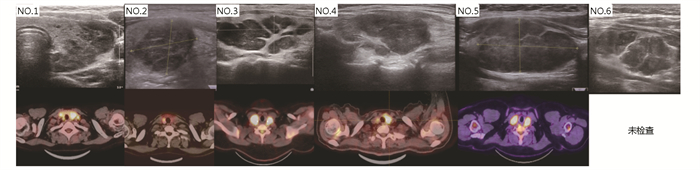
 DownLoad: