| Citation: |
HU Tingting, GUO Lilin, ZHU Yuanyuan, TIAN Zhuang. Imaging Characteristics of Primary Cardiac Angiosarcoma[J]. Journal of Rare Diseases, 2023, 2(1): 88-97. DOI: 10.12376/j.issn.2097-0501.2023.01.012

|
Primary cardiac angiosarcoma is a type of soft tissue sarcoma originating in vascular endothelial cells, without obvious gender differences in the incidence rate and specific early clinical manifestations, whilstpericardial effusion often found at the first presentation of most patients. Tumors are mostly located in the right atrium and pericardium. Echocardiography is the preferred examination method for diagnosing cardiac angiosarcoma and multimodal imaging is important in the diagnosis and differential diagnosis of benign and malignant cardiac mass. This article retrospectively analyzes the 25 cases of clinical manifestations and imaging features of primary cardiac angiosarcoma.
| [1] |
Randhawa JS, Budd GT, Randhawa M, et al. Primary cardiac sarcoma: 25-year cleveland clinic experience[J]. Am J Clin Oncol, 2016, 39(6): 593-599. doi: 10.1097/COC.0000000000000106
|
| [2] |
Florou V, Wilky BA. Current management of angiosarcoma: recent advances and lessons from the past[J]. Curr Treat Options Oncol, 2021, 22(7): 61. doi: 10.1007/s11864-021-00858-9
|
| [3] |
Butany J, Yu W. Cardiac angiosarcoma: two cases and a review of the literature[J]. Can J Cardiol, 2000, 16(2): 197-205.
|
| [4] |
Riles E, Gupta S, Wang DD, et al. Primary cardiac angiosarcoma: a diagnostic challenge in a young man with recurrent pericardial effusions[J]. Exp Clin Cardiol, 2012, 17(1): 39-42.
|
| [5] |
Tang QY, Guo LD, Wang WX, et al. Usefulness of contrast perfusion echocardiography for differential diagnosis of cardiac masses[J]. Ultrasound Med Biol, 2015, 41(9): 2382-2390. doi: 10.1016/j.ultrasmedbio.2015.05.010
|
| [6] |
Chen Y, Li Y, Zhang N, et al. Clinical and imaging features of primary cardiac angiosarcoma[J]. Diagnostics (Basel), 2020, 10(10): 776. doi: 10.3390/diagnostics10100776
|
| [7] |
Li X, Lan L, Hu H. Case report: primary cardiac angiosarcoma with multiple metastases[J]. Front Cardiovasc Med, 2022, 9: 941967. doi: 10.3389/fcvm.2022.941967
|
| [8] |
Yu JF, Cui H, Ji GM, et al. Clinical and imaging manifestations of primary cardiac angiosarcoma[J]. BMC Med Imaging, 2019, 19(1): 16. doi: 10.1186/s12880-019-0318-4
|
| [9] |
Araoz PA, Eklund HE, Welch TJ, et al. CT and MR imaging of primary cardiac malignancies[J]. Radiographics, 1999, 19(6): 1421-1434. doi: 10.1148/radiographics.19.6.g99no031421
|
| [10] |
Göbölös L, Bhatnagar G. Angiosarcoma of the heart[J]. JACC Case Rep, 2021, 3(6): 950-953. doi: 10.1016/j.jaccas.2021.04.030
|
| [11] |
Yin H, Mao W, Tan H, et al. Role of (18)F-FDG PET/CT imaging in cardiac and pericardial masses[J]. J Nucl Cardiol, 2022, 29(3): 1293-1303. doi: 10.1007/s12350-020-02510-9
|
| [12] |
Yaddanapudi K, Brunken R, Tan CD, et al. PET-MR imaging in evaluation of cardiac and paracardiac masses with histopathologic correlation[J]. JACC Cardiovasc Imaging, 2016, 9(1): 82-85. doi: 10.1016/j.jcmg.2015.04.028
|
| [13] |
Bruce CJ. Cardiac tumours: diagnosis and management[J]. Heart, 2011, 97(2): 151-160. doi: 10.1136/hrt.2009.186320
|
| [14] |
Masood I, Duran C, Malik K, et al. Uterineintravenous leiomyomatosis with cardiac involvement[J]. Radiol Case Rep, 2020, 15(8): 1389-1393. doi: 10.1016/j.radcr.2020.05.053
|
| [15] |
Li X, Chen Y, Liu J, et al. Cardiac magnetic resonance imaging of primary cardiac tumors[J]. Quant Imaging Med Surg, 2020, 10(1): 294-313. doi: 10.21037/qims.2019.11.13
|
| [16] |
Kupsky DF, Newman DB, Kumar G, et al. Echocardiographic features of cardiac angiosarcomas: the mayo clinic experience (1976-2013)[J]. Echocardiography, 2016, 33(2): 186-192. doi: 10.1111/echo.13060
|
| [17] |
Hammami MB, Al-Wawi MZ, Fazel H, et al. Incidence, prognostic significance, and survival outcomes of primary cardiac sarcoma: an updated population-based retrospective study[J]. Anatol J Cardiol, 2021, 25(2): 104-110.
|

Official account

Supported by: Beijing Renhe Information Technology Co., Ltd. 
 Author
 Peer Review
 Editorial Entry
 Editor Work
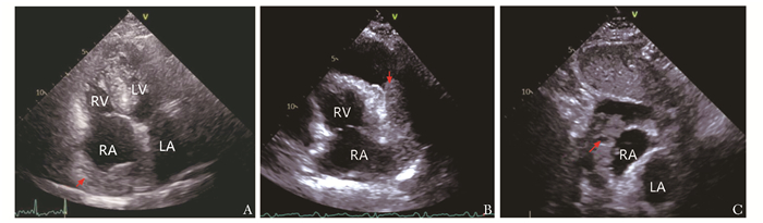
 DownLoad: