| Citation: |
GUO Yubo, WANG Xuezhu, LI Xiao, GAO Yajuan, TIAN Zhuang, LI Jian, HUO Li, WANG Yining. Preliminary Study on Quantitative Evaluation of Myocardial Fibrosis by CardiacMagnetic Resonance in Patients with Light Chain Cardiac Amyloidosis[J]. Journal of Rare Diseases, 2023, 2(1): 43-49. DOI: 10.12376/j.issn.2097-0501.2023.01.006

|
Myocardial fibrosis is a potential mechanism of light-chain myocardial amyloidosis(AL-CA). This research aimed at exploring the correlation between multiparameter cardiac magnetic resonance (CMR) and myocardial fibrosis by relating the CMR myocardial tissue characteristics, the morphological and the functional parameters with gallium-68-labeledfibroblast activation protein inhibitor 04 positron emission tomography (68Ga-FAPI PET).
We gave the patients diagnosed with AL-CA in Peking Union Medical College Hospital from August to December 2021 the examinations of CMR and 68Ga-FAPI PET/CT. We recorded and analyzed the information on clinical manifestations and examinations of the patients.
A total of 23 patients with AL-CA were included, 15 (65.2%)of which were male and the mean age was 58.3±6.5 years. Patients with high 68Ga-FAPI-04 uptake had shown growth in myocardial extracellular volume (ECV), significantly higher than those in the negative group (P=0.047). In addition, patients' myocardial ECV was positively correlated with myocardial FAPI uptake (r=0.628, P=0.001;r=0.727, P < 0.001;r=0.661, P=0.001). Patients in the positive group showd reduced left ventricular (LV) ejection fraction (EF)(P < 0.001).LVEF (r=-0.798, P < 0.001;r=-0.794, P < 0.001; r=-0.795, P < 0.001) and right ventricular (RV)EF (r=-0.735, P < 0.001;r=-0.739, P < 0.001;r=- 0.684, P < 0.001) showd negatively correlated with myocardial FAPI uptake, LV circumferential strain (r=0.668, P < 0.001;r=0.708, P < 0.001;r=0.705, P < 0.001), LV longitudinal strain (r=0.629, P=0.001;r=0.635, P=0.001; r=0.597, P=0.003), and RV longitudinal strain (r=0.575, P=0.004; r=0.792, P < 0.001;r=0.673, P < 0.001) were negatively correlated with myocardial FAPI uptake.
FAPI-related fibroblast activation is concurrent with CMR-related abnormal myocardial interstitial characteristics that leads to the decreased function of the myocardial movement. Patients with increased FAPI uptake present with increased ECV, decreased EF, and decreased strain with morphological abnormalities.
| [1] |
Tillmanns J, Hoffmann D, Habbaba Y, et al. Fibroblast activation protein alpha expression identifies activated fibroblasts after myocardial infarction[J]. J Mol Cell Cardiol, 2015, 87: 194-203. doi: 10.1016/j.yjmcc.2015.08.016
|
| [2] |
Gertz MA, Dispenzieri A. Systemic amyloidosis recognition, prognosis, and therapy: a systematic review[J]. JAMA, 2020, 324(1): 79-89. doi: 10.1001/jama.2020.5493
|
| [3] |
Fontana M, Corović A, Scully P, et al. Myocardial amyloidosis: the exemplar interstitial disease[J]. JACC Cardiovasc Imaging, 2019, 12(11 Pt 2): 2345-2356.
|
| [4] |
Moon JC, Messroghli DR, Kellman P, et al. Myocardial T1 mapping and extracellular volume quantification: a Society for Cardiovascular Magnetic Resonance (SCMR)and CMR Working Group of the European Society of Cardiology consensus statement[J]. J Cardiovasc Magn Reson, 2013, 15(1): 92. doi: 10.1186/1532-429X-15-92
|
| [5] |
Wang X, Guo Y, Gao Y, et al. Feasibility of 68Ga-labeled fibroblast activation protein inhibitor PET/CT in light-chain cardiac amyloidosis[J]. JACC Cardiovasc Imaging, 2022, 15(11): 1960-1970. doi: 10.1016/j.jcmg.2022.06.004
|
| [6] |
Pucci A, Aimo A, Musetti V, et al. Amyloid deposits and fibrosis on left ventricular endomyocardial biopsy correlate with extracellular volume in cardiac amyloidosis[J]. J Am Heart Assoc, 2021, 10(20): e020358. doi: 10.1161/JAHA.120.020358
|
| [7] |
Knight DS, Zumbo G, Barcella W, et al. Cardiac structural and functional consequences of amyloid deposition by cardiac magnetic resonance and echocardiography and their prognostic roles[J]. JACC Cardiovasc Imaging, 2019, 12(5): 823-833. doi: 10.1016/j.jcmg.2018.02.016
|
| [8] |
Mora V, Roldán I, Bertolín J, et al. Influence of ventricular wringing on the preservation of left ventricular ejection fraction in cardiac amyloidosis[J]. J Am Soc Echocardiogr, 2021, 34(7): 767-774. doi: 10.1016/j.echo.2021.02.016
|

Official account

Supported by: Beijing Renhe Information Technology Co., Ltd. 
 Author
 Peer Review
 Editorial Entry
 Editor Work
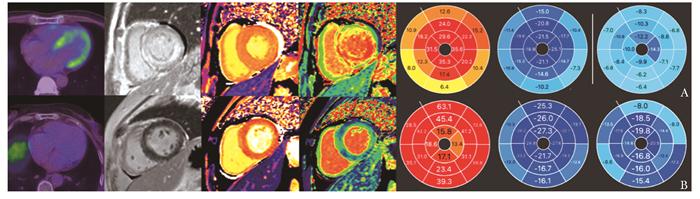
 DownLoad: